Online first
Current issue
Archive
Special Issues
About the Journal
Publication Ethics
Anti-Plagiarism system
Instructions for Authors
Instructions for Reviewers
Editorial Board
Editorial Office
Contact
Reviewers
All Reviewers
2025
2024
2023
2022
2021
2020
2019
2018
2017
2016
General Data Protection Regulation (RODO)
RESEARCH PAPER
Evidence of Babesia microti penetration of hepatocytes based on in vitro and in vivo studies
1
Department of Medical Sciences, Wladyslaw Bieganski Collegium Medicum Jan Dlugosz University, Częstochowa, Poland
2
State Sanitary Inspection, Provincial Sanitary and Epidemiological Station, Katowice, Poland
3
Institute of Biology, Biotechnology and Environmental Protection, Faculty of Natural Sciences, University of Silesia, Katowice, Poland
4
Department of Laryngology, Faculty of Medical Sciences in Katowice, Medical University of Silesia in Katowice, Katowice, Poland
5
Department of Small Livestock Breeding, National Research Institute of Animal Production, Kraków, Poland
6
Department of Biotechnology and Nutrigenomics, Institute of Genetics and Animal Biotechnology of the Polish Academy of Sciences, Jastrzębiec, Poland
Corresponding author
Krzysztof Niemczuk
Biotechnology and Nutrigenomic, Institute of Genetics and Animal Biotechnology of the Polish Academy of Sciences, Postępu, 05-552, Magdalenka, Poland
Biotechnology and Nutrigenomic, Institute of Genetics and Animal Biotechnology of the Polish Academy of Sciences, Postępu, 05-552, Magdalenka, Poland
Ann Agric Environ Med. 2025;32(4):542-551
KEYWORDS
TOPICS
ABSTRACT
Introduction and objective:
Babesiosis is a tick-borne disease of animals and humans caused by the intraerythrocytic protozoa Babesia genus. The objective of this study was to demonstrate that B. microti can invade not only blood cells but also cells of other parenchymal organs, and to examine the effects of this parasitemia. An additional objective was to ascertain whether there were differences in the response of B. microti to hepatocytes in vitro and in vivo conditions.
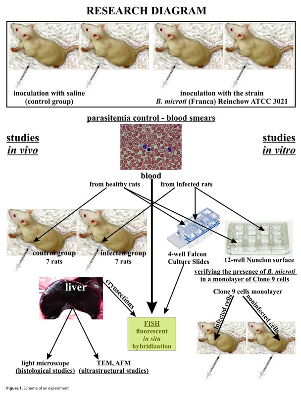
Material and methods:
Wistar rats and the reference hepatocyte cell line Clone 9 isolated from rat livers were used. The rats and cell cultures were infected with an inoculum of B. microti. The investigation of cells in vitro and tissues in vivo was conducted using a light microscope, transmission electron microscope, atomic force microscope, and molecular methods.
Results:
The research findings revealed substantial structural damage to cells, including hydropic degeneration and mitochondrial swelling in B. microti-infected hepatocytes under culture conditions. Similar damage to hepatocytes and thrombosis formation in liver blood vessels were observed. These changes indicated severe liver dysfunction and inflammation of the liver.
Conclusions:
B. microti in intermediate hosts can infect erythrocytes, lymphocytes, and other cells, such as hepatocytes. B. microti affects liver cells directly in in vitro conditions and causes significant liver dysfunction in infected animals. The observed alterations in the blood vessels, including the adhesion of erythrocytes and thrombocytes to the vessel endothelium, provided compelling evidence that the infection of the intermediate host, B. microti, results in haemodynamic disturbances that significantly impact liver dysfunction.
Babesiosis is a tick-borne disease of animals and humans caused by the intraerythrocytic protozoa Babesia genus. The objective of this study was to demonstrate that B. microti can invade not only blood cells but also cells of other parenchymal organs, and to examine the effects of this parasitemia. An additional objective was to ascertain whether there were differences in the response of B. microti to hepatocytes in vitro and in vivo conditions.
Material and methods:
Wistar rats and the reference hepatocyte cell line Clone 9 isolated from rat livers were used. The rats and cell cultures were infected with an inoculum of B. microti. The investigation of cells in vitro and tissues in vivo was conducted using a light microscope, transmission electron microscope, atomic force microscope, and molecular methods.
Results:
The research findings revealed substantial structural damage to cells, including hydropic degeneration and mitochondrial swelling in B. microti-infected hepatocytes under culture conditions. Similar damage to hepatocytes and thrombosis formation in liver blood vessels were observed. These changes indicated severe liver dysfunction and inflammation of the liver.
Conclusions:
B. microti in intermediate hosts can infect erythrocytes, lymphocytes, and other cells, such as hepatocytes. B. microti affects liver cells directly in in vitro conditions and causes significant liver dysfunction in infected animals. The observed alterations in the blood vessels, including the adhesion of erythrocytes and thrombocytes to the vessel endothelium, provided compelling evidence that the infection of the intermediate host, B. microti, results in haemodynamic disturbances that significantly impact liver dysfunction.
REFERENCES (24)
1.
Krause PJ. Human babesiosis. Int J Parasitol. 2019;49(2):165–174. https://doi.org/10.1016/j.ijpa....
2.
Tufts DM, Diuk-Wasser MA. Transplacental transmission of tick-borne Babesia microti in its natural host Peromyscus leucopus. Parasit Vectors. 2018;11(1):286. https://doi.org/10.1186/s13071....
3.
Jasik KP, Okła H, Słodki J, Rozwadowska B, Słodki A, Rupik W. Congenital Tick Borne Diseases: Is This An Alternative Route of Transmission of Tick-Borne Pathogens In Mammals? Vector Borne Zoonotic Dis. 2015;15(11):637–644. https://doi.org/10.1089/vbz.20....
4.
Bloch EM, Krause PJ, Tonnetti L. Preventing Transfusion-Transmitted Babesiosis. Pathogens. 2021;10(9):1176. https://doi.org/10.3390/pathog....
5.
Kumar A, O’Bryan J, Krause PJ. Correction: Kumar, et al. The Global Emergence of Human Babesiosis. Pathogensn2021,n10, 1447. Pathogens. 2022;11(5):607. https://doi.org/10.3390/pathog....
6.
Bajer A, Beck A, Beck R, et al. Babesiosis in Southeastern, Central and Northeastern Europe: An Emerging and Re-Emerging Tick-Borne Disease of Humans and Animals. Microorganisms. 2022;10(5):945. https://doi.org/10.3390/microo....
7.
Moritz ED, Winton CS, Tonnetti L, et al. Screening for Babesia microti in the U.S. Blood Supply. N Engl J Med. 2016;375(23):2236–2245. https://doi.org/10.1056/NEJMoa....
8.
Waked R, Krause PJ. Human Babesiosis. Infect Dis Clin North Am. 2022;36(3):655–670. https://doi.org/10.1016/j.idc.....
9.
Barbiero A, Gabrielli S, Dani L, et al. Babesiosis in the immuno-compro mised population: Results from a multicentric cohort study conducted in Italy. Parasite Epidemiol Control. 2024;26:e00372. https://doi.org/10.1016/j.pare....
10.
Albertyńska M, Okła H, Jasik K, Urbańska-Jasik D, Pol P. Interactions between Babesia microti merozoites and rat kidney cells in a short-term in vitro culture and animal model. Sci Rep. 2021;11(1):23663. https://doi.org/10.1038/s41598....
11.
Hildebrandt A, Gray J, Montero E. Characteristics of Human Babesiosis in Europe. Pathogens. 2023;12(2):323. https://doi.org/10.3390/pathog....
12.
Mazigo E, Jun H, Oh J, et al. Ring stage classification of Babesia microti and Plasmodium falciparum using optical diffraction 3D tomographic technique. Parasit Vectors. 2022;15(1):434. https://doi.org/10.1186/s13071....
13.
Wahl I, Wardemann H. How to induce protective humoral immunity against Plasmodium falciparum circumsporozoite protein. J Exp Med. 2022;219(2):e20201313. https://doi.org/10.1084/jem.20....
14.
Dumic I, Madrid C, Rueda Prada L, Nordstrom CW, Taweesedt PT, Ramanan P. Splenic Complications of Babesia microti Infection in Humans: A Systematic Review. Can J Infect Dis Med Microbiol. 2020;2020:6934149. https://doi.org/10.1155/2020/6....
15.
Capasso C, Supuran CT. The management of Babesia, amoeba and other zoonotic diseases provoked by protozoa. Expert Opin Ther Pat. 2023;33(3):179–192. https://doi.org/10.1080/135437....
16.
Okła H, Jasik KP, Słodki J, et al. Hepatic tissue changes in rats due to chronic invasion of Babesia microti. Folia Biol (Krakow). 2014;62(4):353–359. https://doi.org/10.3409/fb62_4....
17.
Shah JS, Mark O, Caoili E, et al. A Fluorescence in Situ Hybridization (FISH) Test for Diagnosing Babesiosis. Diagnostics (Basel). 2020;10(6):377. https://doi.org/10.3390/diagno....
18.
Goodman A, Bilal M, Amarnath S, Gentile T, Shepherd Z. The unusual case of babesiosis causing disseminated intravascular coagulation with hemophagocytic lymphohistiocytosis. Clin Case Rep. 2021;9(9):e04744. https://doi.org/10.1002/ccr3.4....
19.
Hu Y, Wang M, Ren S, et al. Quantitative proteomics and phosphoproteomic analyses of mouse livers after tick-borne Babesia microti infection. Int J Parasitol. 2021;51(2–3):167–182. https://doi.org/10.1016/j.ijpa....
20.
Uilenberg G. Babesia--a historical overview. Vet Parasitol. 2006;138(1–2):3–10. https://doi.org/10.1016/j.vetp....
21.
Bilić P, Horvatić A, Kuleš J, et al. Serum and urine profiling by high-throughput TMT-based proteomics for the investigation of renal dysfunction in canine babesiosis. J Proteomics. 2023;270:104735. https://doi.org/10.1016/j.jpro....
22.
Khrais A, Ramasamy D, Kumar S. Acalculous Cholecystitis: An Uncommon Presentation of Babesiosis. Cureus. 2022;14(2):e22165. https://doi.org/10.7759/cureus....
23.
Cilekar M, Uysal O, Bal C, Turel S, Yılmaz S. Leptin increases mitotic index and regeneration ratio in hepatectomized rats. Med Sci Monit Basic Res. 2013;19:279–284. https://doi.org/10.12659/MSMBR....
24.
Ha SY, Choi M, Lee T, Park CK. The Prognostic Role of Mitotic Index in Hepatocellular Carcinoma Patients after Curative Hepatectomy. Cancer Res Treat. 2016, 48, 180–189. https://doi.org/10.4143/crt.20....
Share
RELATED ARTICLE
| eISSN: | 1898-2263 |
| ISSN: | 1232-1966 |

Generation of the DOI (Digital Object Identifier) - task financed under the agreement No NrRCN/SP/0532/2021/1 by the Minister of Science and Higher
© 2006-2026 Journal hosting platform by Bentus
We process personal data collected when visiting the website. The function of obtaining information about users and their behavior is carried out by voluntarily entered information in forms and saving cookies in end devices. Data, including cookies, are used to provide services, improve the user experience and to analyze the traffic in accordance with the Privacy policy. Data are also collected and processed by Google Analytics tool (more).
You can change cookies settings in your browser. Restricted use of cookies in the browser configuration may affect some functionalities of the website.
You can change cookies settings in your browser. Restricted use of cookies in the browser configuration may affect some functionalities of the website.