Online first
Current issue
Archive
Special Issues
About the Journal
Publication Ethics
Anti-Plagiarism system
Instructions for Authors
Instructions for Reviewers
Editorial Board
Editorial Office
Contact
Reviewers
All Reviewers
2025
2024
2023
2022
2021
2020
2019
2018
2017
2016
General Data Protection Regulation (RODO)
REVIEW PAPER
Rapid, Reproducible and Reliable – multimodal AI for CT, chest X-ray and clinical data integration with insights from COVID-19
1
Department of Correct, Clinical and Imaging Anatomy, Medical University, Lublin, Poland
2
Euroimmun DNA, Wrocław, Poland
3
Medicos Medical Centre, Lublin, Poland
4
Department of Radiology and Nuclear Medicine, Medical University, Lublin, Poland
These authors had equal contribution to this work
Corresponding author
Karolina Kołodziejczyk
Department of Correct, Clinical and Imaging Anatomy, Medical University of Lublin, Poland
Department of Correct, Clinical and Imaging Anatomy, Medical University of Lublin, Poland
KEYWORDS
COVID-19chest X-raydeep learningchest computed tomographymultimodal artificial intelligenceclinical datalaboratory dataseverity predictionclinical decision supportalgorithmic fairness
TOPICS
ABSTRACT
Introduction and objective:
The COVID-19 pandemic accelerated development of multimodal artificial intelligence (AI) models that combine chest computed tomography (CT), chest X-ray (CXR) and clinical/laboratory data to support imaging-based diagnosis, triage and prognostication. This review synthesizes reported performance, clinical utility and limitations.
Review methods:
PubMed, Scopus and Google Scholar (Jan 2020–Jun 2025) were searched and included peer-reviewed English studies applying machine learning or deep learning to CT and/or CXR with reported sample sizes and performance metrics (AUC, sensitivity, specificity, F1). Preprints, case reports and studies without sample sizes or performance metrics were excluded.
Brief description of the state of knowledge:
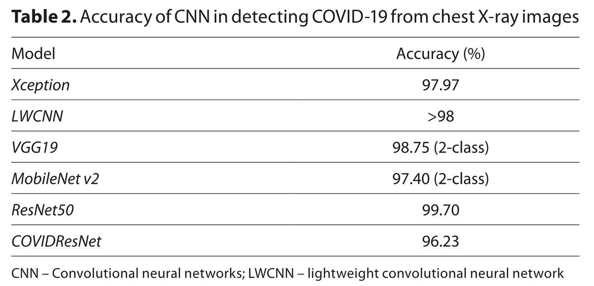
Meta-analyses report high discriminative performance for severity prediction (pooled AUC ≈ 0.89) alongside a high prevalence of study bias. Selected studies reported top accuracies up to ≈98% and multimodal F1 scores up to 0.89. Recurring limitations were dataset heterogeneity, single-centre training and scarce external validation. Some reports found automated pipelines substantially faster than manual reads (e.g. ~2.7 s vs ~6.5 min), although workflow times vary by setting.
Summary:
Multimodal integration of CT, CXR and clinical data with AI is promising for rapid, reproducible assessment of COVID-19 severity. Clinical translation requires standardized acquisition and reporting, rigorous multicentre external validation, transparent methods, and formal evaluation of clinical impact and fairness.
The COVID-19 pandemic accelerated development of multimodal artificial intelligence (AI) models that combine chest computed tomography (CT), chest X-ray (CXR) and clinical/laboratory data to support imaging-based diagnosis, triage and prognostication. This review synthesizes reported performance, clinical utility and limitations.
Review methods:
PubMed, Scopus and Google Scholar (Jan 2020–Jun 2025) were searched and included peer-reviewed English studies applying machine learning or deep learning to CT and/or CXR with reported sample sizes and performance metrics (AUC, sensitivity, specificity, F1). Preprints, case reports and studies without sample sizes or performance metrics were excluded.
Brief description of the state of knowledge:
Meta-analyses report high discriminative performance for severity prediction (pooled AUC ≈ 0.89) alongside a high prevalence of study bias. Selected studies reported top accuracies up to ≈98% and multimodal F1 scores up to 0.89. Recurring limitations were dataset heterogeneity, single-centre training and scarce external validation. Some reports found automated pipelines substantially faster than manual reads (e.g. ~2.7 s vs ~6.5 min), although workflow times vary by setting.
Summary:
Multimodal integration of CT, CXR and clinical data with AI is promising for rapid, reproducible assessment of COVID-19 severity. Clinical translation requires standardized acquisition and reporting, rigorous multicentre external validation, transparent methods, and formal evaluation of clinical impact and fairness.
REFERENCES (42)
1.
Becker J, Decker JA, Römmele C, et al. Artificial Intelligence-Based Detection of Pneumonia in Chest Radiographs. Diagnostics (Basel). 2022;12(6):1465. doi:10.3390/diagnostics12061465.
2.
Quanyang W, Yao H, Sicong W, et al. Artificial intelligence in lung cancer screening: Detection, classification, prediction, and prognosis. Cancer Med. 2024;13(7):e7140. doi:10.1002/cam4.7140.
3.
Viswanathan VS, Toro P, Corredor G, et al. The state of the art for artificial intelligence in lung digital pathology. J Pathol. 2022;257(4):413–429. doi:10.1002/path.5966.
4.
Aboshosha A. AI based medical imagery diagnosis for COVID-19 disease examination and remedy. Sci Rep. 2025;15(1):1607. doi:10.1038/s41598-024-84644-1.
5.
Alsharif W, Qurashi A. Effectiveness of COVID-19 diagnosis and management tools: A review. Radiography (Lond). 2021;27(2):682–687. doi:10.1016/j.radi.2020.09.010.
6.
Gkekas I, Katsamakas S, Mylonas S, et al. AI Promoted Virtual Screening, Structure-Based Hit Optimization, and Synthesis of Novel COVID-19 S-RBD Domain Inhibitors. J Chem Inf Model. 2024;64(22):8562–8585. doi:10.1021/acs.jcim.4c01110.
7.
Kosar A, Asif M, Ahmad MB, et al. Towards classification and comprehensive analysis of AI-based COVID-19 diagnostic techniques: A survey. Artif Intell Med. 2024;151:102858. doi:10.1016/j.artmed.2024.102858.
8.
Wang D, Hu B, Hu C, et al. Clinical Characteristics of 138 Hospitalized Patients With 2019 Novel Coronavirus-Infected Pneumonia in Wuhan, China. JAMA. 2020;323(11):1061–1069. doi:10.1001/jama.2020.1585.
9.
LeBlanc JJ, Heinstein C, MacDonald J, et al. A combined oropharyngeal/nares swab is a suitable alternative to nasopharyngeal swabs for the detection of SARS-CoV-2. J Clin Virol. 2020;128:104442. doi:10.1016/j.jcv.2020.104442.
10.
Topff L, Ranschaert ER, Bartels-Rutten A, et al. Artificial Intelligence Tool for Detection and Worklist Prioritization Reduces Time to Diagnosis of Incidental Pulmonary Embolism at CT. Radiol Cardiothorac Imaging. 2023;5(2):e220163. doi:10.1148/ryct.220163.
11.
Catalano M, Bortolotto C, Nicora G, et al. Performance of an AI algorithm during the different phases of the COVID pandemics: what can we learn from the AI and vice versa. Eur J Radiol Open. 2023;11:100497. doi:10.1016/j.ejro.2023.100497.
12.
Górecki A, Piech P, Kołodziejczyk K, et al. Synergistic Imaging: Combined Lung Ultrasound and Low-Dose Chest CT for Quantitative Assessment of COVID-19 Severity—A Prospective Observational Study. Diagnostics. 2025;15(15):1875. doi:10.3390/diagnostics15151875.
13.
Wang S, Kang B, Ma J, et al. A deep learning algorithm using CT images to screen for Coronavirus disease (COVID-19). Eur Radiol. 2021;31(8):6096–6104. doi:10.1007/s00330-021-07715-1.
14.
Moezzi M, Shirbandi K, Shahvandi HK, et al. The diagnostic accuracy of Artificial Intelligence-Assisted CT imaging in COVID-19 disease: A systematic review and meta-analysis. Inform Med Unlocked. 2021;24:100591. doi:10.1016/j.imu.2021.100591.
15.
Ardakani AA, Kanafi AR, Acharya UR, et al. Application of deep learning technique to manage COVID-19 in routine clinical practice using CT images: Results of 10 convolutional neural networks. Comput Biol Med. 2020;121:103795. doi:10.1016/j.compbiomed.2020.103795.
16.
Karpiel I, Starcevic A, Urzeniczok M. Database and AI Diagnostic Tools Improve Understanding of Lung Damage, Correlation of Pulmonary Disease and Brain Damage in COVID-19. Sensors (Basel). 2022;22(16):6312. doi:10.3390/s22166312.
17.
Javor D, Kaplan H, Kaplan A, et al. Deep learning analysis provides accurate COVID-19 diagnosis on chest computed tomography. Eur J Radiol. 2020;133:109402. doi:10.1016/j.ejrad.2020.109402.
18.
Shen D, Wu G, Suk HI. Deep Learning in Medical Image Analysis. Annu Rev Biomed Eng. 2017;19:221–248. doi:10.1146/annurev-bioeng-071516-044442.
19.
Singh D, Kumar V, Vaishali, et al. Classification of COVID-19 patients from chest CT images using multi-objective differential evolution-based convolutional neural networks. Eur J Clin Microbiol Infect Dis. 2020;39(7):1379–1389. doi:10.1007/s10096-020-03901-z.
20.
Shan F, Gao Y, Wang J, et al. Abnormal lung quantification in chest CT images of COVID-19 patients with deep learning and its application to severity prediction. Med Phys. 2021;48(4):1633–1645. doi:10.1002/mp.14609.
21.
Górecki A, Piech P, Bronikowska A, et al. Safe, Smart, and Scalable: A Prospective Multicenter Study on Low-Dose CT and CTSS for Emergency Risk Stratification in COVID-19. J Clin Med. 2025;14(13):4423. doi:10.3390/jcm14134423.
22.
Apostolopoulos ID, Mpesiana TA. Covid-19: automatic detection from X-ray images utilizing transfer learning with convolutional neural networks. Phys Eng Sci Med. 2020;43(2):635–640. doi:10.1007/s13246-020-00865-4.
23.
Narin A, Kaya C, Pamuk Z. Automatic detection of coronavirus disease (COVID-19) using X-ray images and deep convolutional neural networks. Pattern Anal Appl. 2021;24(3):1207–1220. doi:10.1007/s10044-021-00984-y.
24.
Farooq M, Hafeez A. Covid-resnet: A deep learning framework for screening of covid19 from radiographs. arXiv. 2020. 2003.14395.
25.
Wang L, Lin ZQ, Wong A. COVID-Net: a tailored deep convolutional neural network design for detection of COVID-19 cases from chest X-ray images. Sci Rep. 2020;10(1):19549. doi:10.1038/s41598-020-76550-z.
26.
Ucar F, Korkmaz D. COVIDiagnosis-Net: Deep Bayes-SqueezeNet based diagnosis of the coronavirus disease 2019 (COVID-19) from X-ray images. Med Hypotheses. 2020;140:109761. doi:10.1016/j.mehy.2020.109761.
27.
Ozturk T, Talo M, Yildirim EA, et al. Automated detection of COVID-19 cases using deep neural networks with X-ray images. Comput Biol Med. 2020;121:103792. doi:10.1016/j.compbiomed.2020.103792.
28.
Tenda ED, Yunus RE, Zulkarnaen B, et al. Comparison of the discrimination performance of AI scoring and the Brixia score in predicting COVID19 severity on chest X-ray imaging: diagnostic accuracy study. JMIR Form Res. 2024;8:e46817. doi:10.2196/46817.
29.
Yang Y, Zhang H, Gichoya JW, et al. The limits of fair medical imaging AI in real-world generalization. Nat Med. 2024;30(10):2838–2848. doi:10.1038/s41591-024-031134.
30.
Jackson NJ, Yan C, Malin BA. Enhancement of fairness in AI for chest X-ray classification. AMIA Annu Symp Proc. 2024;2024:551–560.
31.
Rakovics M, Meznerics FA, Fehérvári P, et al. Deep neural networks excel in COVID-19 disease severity prediction—metaregression analysis. Sci Rep. 2025;15(1):10350. doi:10.1038/s41598-025-952826.
32.
Giełczyk A, Marciniak A, Tarczewska M, et al. Preprocessing methods in chest X-ray image classification. PLoS One. 2022;17(4):e0265949. doi:10.1371/journal.pone.0265949.
33.
Verma K, Sikka G, Swaraj A, et al. Classification of COVID-19 on chest X-ray images using deep learning model with histogram equalization and lung segmentation. SN Comput Sci. 2024;5(4):379. doi:10.1007/s42979-024-026957.
34.
Rahman T, Chowdhury MEH, Khandakar A, et al. BIOCXRNET: a robust multimodal stacking machine learning technique for mortality risk prediction of COVID-19 patients using chest X-ray images and clinical data. Neural Comput Appl. 2023;35:17461–17483. doi:10.1007/s00521-023-08606-w.
35.
Dipaola F, Gatti M, Giaj Levra A, et al. Multimodal deep learning for COVID-19 prognosis prediction in the emergency department: a bicentric study. Sci Rep. 2023;13(1):10868. doi:10.1038/s41598-023-375123.
36.
Jin C, Chen W, Cao Y, et al. Development and evaluation of an artificial intelligence system for COVID-19 diagnosis. Nat Commun. 2020;11(1):5088. doi:10.1038/s41467-020-18685-1.
37.
Hemdan EED, Shouman MA, Karar ME. Covidx-net: A framework of deep learning classifiers to diagnose covid-19 in x-ray images. arXiv. 2020. 2003.11055.
38.
Abbas A, Abdelsamea MM, Gaber MM. Classification of COVID-19 in chest X-ray images using DeTraC deep convolutional neural network. Appl Intell (Dordr). 2021;51(2):854–864. doi:10.1007/s10489-020-01829-7.
39.
Gulakala R, Markert B, Stoffel M. Rapid diagnosis of Covid-19 infections by a progressively growing GAN and CNN optimisation. Comput Methods Programs Biomed. 2023;229:107262. doi:10.1016/j.cmpb.2022.107262.
40.
Waheed A, Goyal M, Gupta D, et al. CovidGAN: Data Augmentation Using Auxiliary Classifier GAN for Improved Covid-19 Detection. IEEE Access. 2020;8:91916–91923. doi:10.1109/ACCESS.2020.2994762.
41.
Liu R, Han H, Liu F, et al. Positive rate of RT-PCR detection of SARS-CoV-2 infection in 4880 cases from one hospital in Wuhan, China, from Jan to Feb 2020. Clin Chim Acta. 2020;505:172–175. doi:10.1016/j.cca.2020.03.009.
42.
Chu DKW, Pan Y, Cheng SMS, et al. Molecular Diagnosis of a Novel Coronavirus (2019-nCoV) Causing an Outbreak of Pneumonia. Clin Chem. 2020;66(4):549–555. doi:10.1093/clinchem/hvaa029.
Share
RELATED ARTICLE
| eISSN: | 1898-2263 |
| ISSN: | 1232-1966 |

Generation of the DOI (Digital Object Identifier) - task financed under the agreement No NrRCN/SP/0532/2021/1 by the Minister of Science and Higher
© 2006-2026 Journal hosting platform by Bentus
We process personal data collected when visiting the website. The function of obtaining information about users and their behavior is carried out by voluntarily entered information in forms and saving cookies in end devices. Data, including cookies, are used to provide services, improve the user experience and to analyze the traffic in accordance with the Privacy policy. Data are also collected and processed by Google Analytics tool (more).
You can change cookies settings in your browser. Restricted use of cookies in the browser configuration may affect some functionalities of the website.
You can change cookies settings in your browser. Restricted use of cookies in the browser configuration may affect some functionalities of the website.